Study Overview
Researchers at Guangxi Medical University Hospital in China have reported promising mid-term results using patient-matched, 3D printed titanium prostheses combined with mesh patches to reconstruct the distal radius after en bloc resection of aggressive giant cell tumors (GCT). The small retrospective series, five patients treated between January 2018 and January 2021, shows that carefully designed metal implants can deliver useful wrist function, support early rehabilitation, and keep complication rates low in the early-to-mid postoperative period.
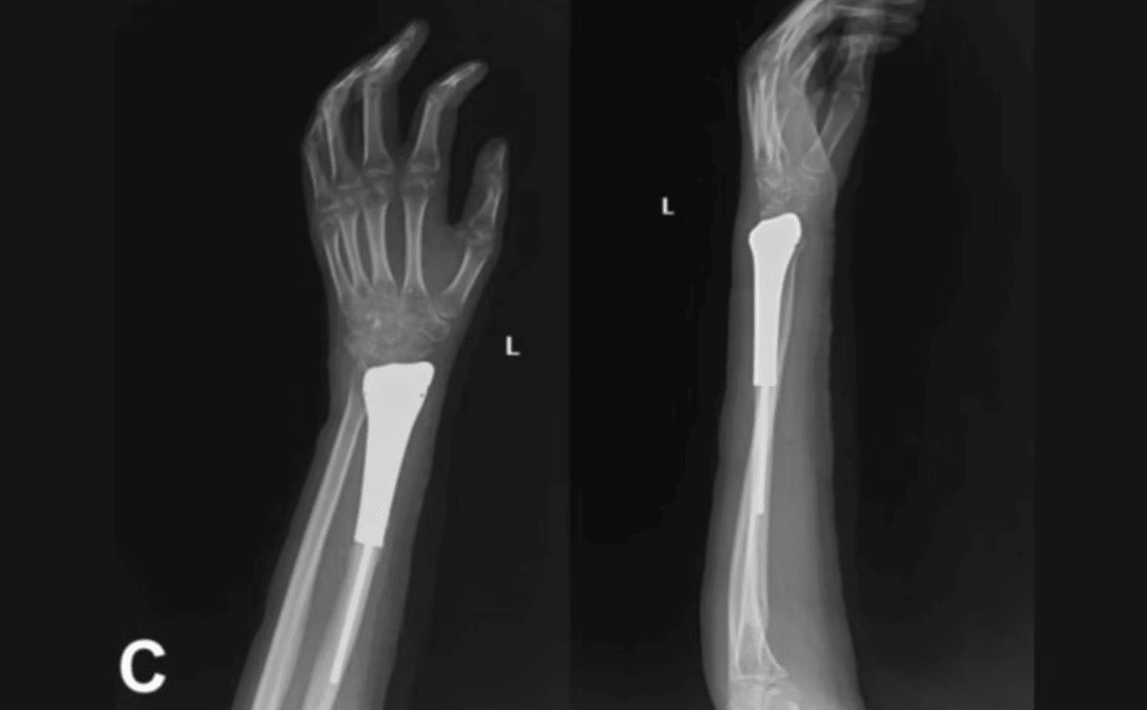
Image Source: VoxelMatters
Patient-Matched Implant Design and Soft-Tissue Reconstruction
The study team designed fully integrated titanium alloy prostheses from each patient’s CT scans and manufactured the implants with an industry partner. Key design features included a textured stem for improved bone-cement grip, a porous trabecular collar to encourage bone bridging, and prefabricated suture holes on the distal prosthetic surface to reattach ligaments and soft tissue. A two-layer PTFE mesh patch was wrapped and sutured to the prosthesis to create a soft-tissue envelope and stabilize the reconstruction, enabling earlier active motion. The authors outline a reproducible intraoperative workflow for prosthesis placement, ligament repair, and cemented stem fixation.
Mid-Term Functional Outcomes and Complications
Functional outcomes were assessed over an average follow-up of 40.8 months (range 32–66 months). At the last follow-up, the mean wrist range of motion on the affected side measured roughly 20° extension and 21.6° flexion, with pronation around 71.2° and supination about 50°; mean grip strength on the treated side was 64.2% of the contralateral hand. The average Mayo wrist score reported was 70, indicating fair to good function. Importantly, the cohort experienced no aseptic loosening, prosthesis infections, or radiocarpal subluxation. There were two cases of distal radioulnar joint (DRUJ) dislocation, and one patient developed ulnar impaction changes by 12 months. No tumor recurrences or metastases were observed during the follow-up window.
What This Means for Clinicians and 3D Printing Providers
The study illustrates how additive manufacturing enables anatomically accurate, multi-feature implants that integrate fixation, soft-tissue attachment points, and porous regions for biological integration, all tailored from the patient’s imaging. The concurrent use of mesh patches to reconstruct a soft-tissue sleeve appears to facilitate early postoperative exercise, which the authors link to faster functional recovery. For 3D printing service bureaus and medical device teams, the case series highlights the importance of close surgeon-engineer collaboration, tight tolerances on articular geometry, and careful post-print finishing to meet surgical performance goals.
Limitations and Future Directions
It is a small retrospective series with mid-term follow-up. Larger, comparative studies and longer surveillance are needed to confirm durability, degenerative outcomes, and broader safety. Additional work should also track functional trajectories against alternative reconstructions and explore refinements in mesh materials, porous gradient design, and fixation strategies. Still, the paper contributes to growing evidence that custom metal 3D printing prostheses, when paired with thoughtful soft-tissue reconstruction, can be a viable reconstructive option for high-demand joints such as the wrist.
Sources: Original open-access study from Guangxi Medical University Hospital (3D Print Med, 2025) and technical news coverage summarized on VoxelMatters.
 0
0