When stranded California sea lions begin washing ashore in large numbers, rapid and accurate diagnostics can make the difference between recovery and fatality. Recently, researchers developed a highly realistic 3D printed pelvis model designed specifically to help veterinarians practice blood collection procedures on these marine mammals. The innovation demonstrates how additive manufacturing is extending beyond traditional medical and industrial applications into wildlife rescue and conservation.
According to reporting by VoxelMatters, the project centers on creating an anatomically accurate training phantom that allows clinicians to safely rehearse needle placement and vascular access techniques. The model was designed to replicate the pelvic anatomy of California sea lions, providing both visual and tactile realism. It is especially important as harmful algal blooms and domoic acid toxicity events continue to impact marine mammals along the U.S. West Coast, increasing the demand for efficient triage and diagnostic procedures.
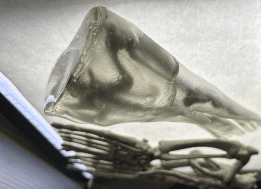
Image Source: John Domol
From CT Data to Physical Model
The research team, based at the University of Nevada, Las Vegas, began by working with high-resolution CT scan data. Using DICOM medical imaging files, they segmented the pelvis into distinct anatomical components. This digital workflow enabled precise reconstruction of skeletal geometry, soft tissue regions, and external contours.
From there, the team employed a multi-material fabrication strategy. Rigid 3D printing technologies were used to produce the skeletal core with accurate structural detail. To simulate muscle and blubber layers, researchers incorporated gelatin-like materials selected for their viscoelastic properties. Finally, a flexible outer shell was fabricated to mimic skin and provide realistic resistance during needle insertion.
The result is a layered phantom that allows trainees not only to see anatomical landmarks but also to feel realistic tissue feedback during procedures. Unlike static teaching aids, this model replicates procedural dynamics, including the resistance encountered when advancing a needle through soft tissue.
The peer-reviewed findings describing this fabrication workflow were published in Scientific Reports, highlighting the interdisciplinary combination of medical imaging, materials science, and 3D printing.
Why This Matters for Marine Mammal Rescue
Veterinary teams responding to stranded sea lions often work under time-sensitive and high-pressure conditions. Blood collection from the caudal gluteal region is a critical diagnostic step used to assess toxicity, infection, and overall health status. However, performing this procedure on a live, stressed animal presents challenges, both technically and ethically.
Traditionally, hands-on training opportunities may rely on cadavers or supervised live procedures. Both options come with limitations, including availability, ethical concerns, and variability in anatomy. A reusable, anatomically precise phantom offers several key advantages:
• Improved clinician confidence: Repeated practice in a controlled environment allows practitioners to refine technique before working with live animals.
• Reduced animal stress: Faster, more accurate needle placement can minimize handling time.
• Standardized training: Multiple trainees can practice on identical models, ensuring consistent educational outcomes.
By reducing reliance on opportunistic training materials, 3D printing introduces a scalable solution that can be distributed across marine mammal rescue centers and veterinary programs.
The Broader Implications of Anatomical 3D Printing
While this project focuses on California sea lions, the workflow is adaptable to other species and procedures. Any scenario involving CT or MRI imaging data can potentially be converted into a custom training model. It opens doors for:
• Wildlife conservation training programs
• Exotic animal veterinary education
• Pre-surgical rehearsal models
• Procedure-specific simulation tools
The combination of DICOM segmentation, rigid and flexible material printing, and gel casting demonstrates how additive manufacturing can replicate both structural and mechanical properties of biological tissues. As materials continue to evolve, we can expect even more lifelike training simulators across the veterinary and human healthcare fields.
Sources
1. VoxelMatters, 3D-printed pelvis model aids diagnostics in beached sea lions.
https://www.voxelmatters.com/3d-printed-pelvis-model-diagnostics-beached-sea-lions/
2. Scientific Reports, peer-reviewed study describing the CT-derived phantom and fabrication methods.
https://www.nature.com/articles/s41598-026-36154-5
3. University of Nevada, Las Vegas, research team and lab responsible for the phantom development.
 0
0


COMMENTS
- Be the first to share your thoughts!